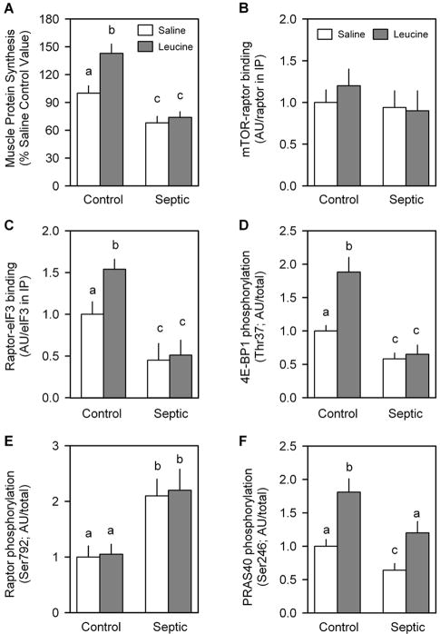
Fig. 9.
Leucine-induced changes in protein synthesis and mTORC1 in skeletal muscle from control and septic rats. Gastrocnemius was sampled 30 min after oral administration of a maximally stimulating dose of the branched-chain amino acid leucine. In vivo muscle protein synthesis was determined as described in Materials and Methods (A). Bar graphs are quantitation of immunoblots after immunoprecipitation (IP) of either raptor (B) or eIF3b (C). Western blot data from whole muscle tissue homogenates have also been quantitated (D, E, and F). There was no sepsis or leucine effect on the total amount of raptor, mTOR, eIF3b, 4E-BP1, or PRAS40 (data not shown). Saline-treated control values were arbitrarily set to 1.0 AU. Values are means ± SEM; n = 7–9 rats per group. Values with different letters are statistically different (P < 0.05).

