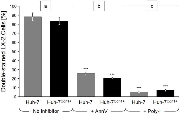
Fig. 1.
Uptake of apoptotic bodies (ABs) by LX-2 cells. LX-2 cells were labeled with green fluorescence and then incubated with red fluorescent ABs for 4 h. ABs were generated from Huh-7 cells or Con1-positive Huh-7 cells by UV-irradiation (100 mJ/cm2) for 24 h, followed by red fluorescence labeling. Numbers of total and double-stained cells were determined by fluorescence microscopy. (a) Without inhibitors; (b) After PS masking with 10 μg/ml annexin-V (AnnV); or (c) incubation with the scavenger receptor antagonist, poly-inosine (Poly-I) at 100 μg/ml. Bars depict percentages of double-stained cells ± standard deviations. Conditions with vs. without inhibitors: ***: p < 0.0001.

