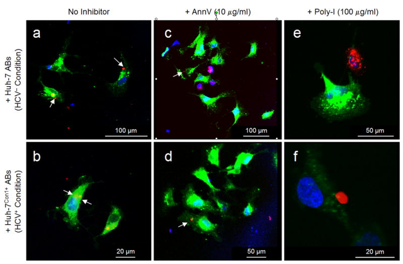
Fig. 2.

LX-2 cells ingest HCV− and HCV+ ABs. Representative photomicrographs depict green fluorescent cells that internalized red fluorescent ABs of HCV− Huh-7 cells (a, c, e) or HVC+ Huh-7Con1+ cells (b, d, f) upon incubation with ABs for 2 h, as visualized by confocal LASER scanning microscopy. Nuclei were counterstained with DAPI (blue). Cells were kept without inhibitors (a, b), or were incubated with AnnV (c, d) or Poly-I (e, f), respectively (cf. Fig. 1 for further detail). White arrows point at ABs (red) inside LX-2 cells (green). [Excitation wave lengths vs. detection ranges: blue fluorescence (λ = 364 nm vs. λ = 385–470 nm); green fluorescence (λ = 488 nm vs. λ = 505–530 nm); and red fluorescence (λ = 543 nm vs. λ = 560 nm)].
