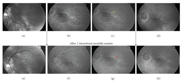
Figure 1.
(a), (b), (c), (d): imaging before intravitreal bevacizumab. (e), (f), (g), (h): imaging after intravitreal bevacizumab. Red free picture (a) shows tilting of optic disk with inferior staphyloma. RPE pigmentary changes are evident along the staphyloma area. In early mid-phases of fluorescein angiography (b, c) an hyperfluorescent dot (yellow arrow) becomes evident over a background of diffuse oblique-oriented RPE atrophy that typically overlies the staphyloma. At 5-minutes late phase (d) leaking point intensity dissolves into a diffuse vanishing hyperfluorescence. After treatment the primitive leaking dot (red arrow) is not detectable in early (f), middle (g), or late phases (h) of fluorangiography.

