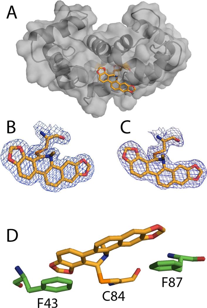
Figure 10.
The x-ray structure of the SC0844-Ca2+-S100B complex. (A) Ribbon and surface diagram of sanguinarine (SC0844)-Ca2+-S100B, illustrating the location of SC0844 (SC0844, orange; nitrogen, blue; oxygen, red; sulfur, orange) covalently bound to Cys84. (B and C) The electron density maps calculated with the 2mFo-DFc coefficients (1.0σ) for SC0833 in each monomer of the biological dimer. (D) Residues in Ca2+-S100B involved in coordinating SC0844. (Protein Databank accession number: 3LLE)

