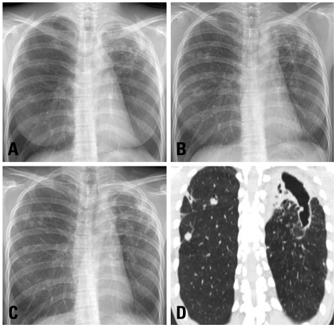
Fig. 1.
A 35 year-old woman with Mycobacterium celatum lung disease. (A) The posteroanterior chest radiography at first visit shows a large cavitary lesion in the left upper lobe. Note the multiple nodular opacities involving the right lung. (B) The chest radiography at the subsequent revisit shows aggravation of the cavitation in the left upper lobe, as well as enlargement of the nodular lesions in the right lung. (C) The chest radiography taken after 12 months of antibiotic treatment shows that the size of the cavitary lesion in the left upper lobe is unchanged, although the multiple nodular lesions in the right lung have improved. (D) The chest computed tomography scan taken after 12 months of antibiotic treatment reveals a large remnant cavitary lesion that involves both the left upper lobe and the superior segment of the left lower lobe.

