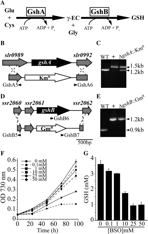
Figure 1.
Disruption of glutathione biosynthesis in Synechocystis 6803. A, Diagram of the glutathione biosynthetic pathway. B, The entire open reading frame of the gshA gene was replaced with a kanamycin resistance cassette (KmR) to generate the ΔgshA::KmR strain. C, Segregation of ΔgshA::KmR was tested by PCR using primers GshA5 and GshA6 shown in A. Lanes show wild-type genomic DNA (WT), pSL2083 (+), and ΔgshA::KmR genomic DNA. D, The gshB gene was replaced with a gentamicin resistance cassette (GmR). E, Segregation of ΔgshB::GmR was confirmed by PCR using primers GshB5, GshB6, and GshB7 shown in D. Lanes show wild-type genomic DNA (WT), pSL2085 (+), and ΔgshB::GmR genomic DNA. F, Growth of wild-type cells in the presence of the GshA inhibitor BSO. G, Cellular GSH concentration after 96 h of growth in the presence of BSO. Primer sequences used in cloning and segregation analysis are shown in Table I.

