Figure 1.
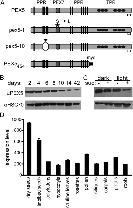
PEX5 gene expression and protein concentration. A, Cartoon representation of the PEX5 protein, the pex5 mutant proteins, and the PEX5454 truncation. The pex5-1 Ser-to-Leu point mutation (Zolman et al., 2000) and the pex5-10 insertion site and resulting deletion are indicated by a star and a triangle, respectively. Gray circles indicate the PEX7-binding domain (PEX7), black ovals indicate the TPR domains, and rectangles represent the PPR domains. The goalpost under the protein indicates the recognition site of the PEX5 antibody (Zolman and Bartel, 2004). B, Total protein was extracted from wild-type Col-0 seedlings and adult plants and analyzed by western blot for PEX5 levels. C, Total protein from 5-d-old wild-type seedlings grown in the dark or light in the presence or absence of Suc was extracted and analyzed by western blot for PEX5 levels. For both B and C, equal amounts of protein were loaded, as confirmed by immunoblotting with an HSC70 antibody (bottom). D, Levels of PEX5 mRNA expression (absolute values) in dry and imbibed seeds, cotyledons, hypocotyls, cauline leaves, rosette leaves, mature pollen, seeds/siliques, carpels, petals, and roots. The graph was constructed using Arabidopsis eFP Browser data from http://bar.utoronto.ca/efp/cgi-bin/efpWeb.cgi (Winter et al., 2007). Data were retrieved on March 6, 2010.

