Figure 6.
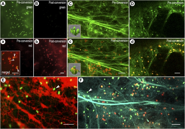
Visualizing motile organelles using mEosFP alone and in combination with other FPs. A, a, B, and b, Mito-mEosFP transiently expressed in tobacco leaves by means of agroinfiltration efficiently targets to mitochondria (A) and is near fully photoconverted (b) after a 30-s exposure to 365/50-nm wavelength. Microscope system 4 was used for acquiring these images. Note that while preconversion fluorescence is barely detectable in the red channel (a) following photoconversion, it is not picked up in the green channel (B). The exposure times for images A and B are identical, as are those for images a and b. The inset in a is a merged image of the green and red channels postconversion using identical exposure times and demonstrates the slight shift and resultant artifact (arrowheads) that occurred during the time lapse between capture of the two sequential images, which could be misinterpreted as an absence of colocalization. C and c, Golgi bodies highlighted by mEosFP::GONST1 and F-actin (highlighted using GFP::mTalin) covisualized before (C) and after photoconversion (c). The rapid motility of Golgi bodies does not allow direct comparisons to be drawn between preconversion and postconversion images in separate channels; therefore, merged images acquired in both channels are presented. The inset three-dimensional surface plot clearly shows quantifiable changes in the color of Golgi bodies from their prephotoconversion to their postphotoconversion state (for more details, see Supplemental Fig. S2). D and d, Peroxisomes labeled with mEosFP-PTS1 and F-actin labeled using YFP::mTalin covisualized before (D) and after photoconversion (d). E, Peroxisomes labeled with mEosFP-PTS1 can be clearly discriminated (arrowhead) from RFP-labeled ER during prolonged covisualization using 488- and 543-nm lasers. Unintended photoconversion of mEosFP does not occur. F, Both the nonphotoconverted (arrowhead, green) and photoconverted (circle, red) forms of mEosFP-PTS1 covisualized with CFP::mTalin targeted to F-actin. Photoconversion was carried out separately, since the 458-nm argon laser line does not cause mEosFP to change color. Images in C to F were acquired using microscope system 2. Bars = 5 μm.

