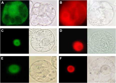
Figure 3.
Subcellular localization of BEIL1 and BERF1 in transiently transformed tobacco protoplasts. A and B, Cytoplasmic and nuclear localization of GFP and RFP, respectively. C and D, Nuclear localization of BEIL1 fused to GFP and RFP, respectively. E and F, Nuclear localization of BERF1 fused to GFP and RFP, respectively. To the right of each image, the corresponding bright-field image is shown.

