Figure 7.

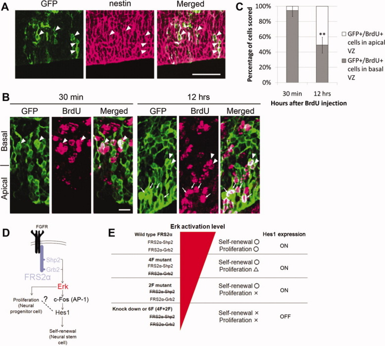
Cells expressing Hes1 show radial glial properties (A–C). (A, B): Vectors expressing Hes1, GFP, and shRNA for FRS2α were coinjected into the lateral ventricles of E13.5 mouse embryos in utero and electroporated. Then, the embryos were sacrificed at E15.5, and cortical sections were immunostained against GFP (green) and nestin (magenta; [A]) or BrdU (magenta; [B]). BrdU was administered 30 minutes or 12 hours before sacrifice. (A): Most GFP-positive cells expressed nestin, as indicated by arrowheads. Scale bar = 50 μm. (B): The cell bodies of most GFP-positive cells in the S-phase (30 minutes after BrdU administration) were located at the basal half of VZ (arrowhead). In contrast, the cell bodies of GFP-positive cells in the G1 phase were present at both the basal and apical (arrow) half of VZ. Scale bar = 10 μm. (C): Quantification of the data in (B). In each condition, at least 145 cells were counted in 19–29 randomly selected areas. (D): Fibroblast growth factor (FGF)-induced Erk activation via Shp2- or Grb2-binding sites of FRS2α contributes to both proliferation of neural stem/progenitor cells (NSPCs) and self-renewal of neural stem cells (NSCs). The latter is at least partly mediated by Hes1, whose expression may be induced by the binding of AP-1 complex to the Hes1 promoter. (E): For the normal proliferation of NSPCs in response to FGF, strong Erk activation via both Shp2- and Grb2-binding sites of FRS2α is required. On the other hand, relatively weak Erk activation levels are sufficient to activate the self-renewal switch of NSCs with Hes1 expression, the master regulator for stemness. Abbreviations: BrdU, Bromodeoxyuridine; FGFR, Fibroblast growth factor receptor; FRS2α, FGF receptor substrate 2α; GFP, Green Fluorescent Protein; VZ, ventricular zone.
