
Figure 1.
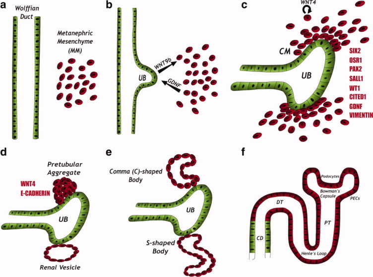
Kidney development. (A): The kidney is formed via reciprocal interactions between two precursor tissues derived form the intermediate mesoderm: the Wolffian duct and the MM. (B): MM-derived signals, mainly the glial-derived neurotrphic factor, induce an outgrowth from the Wolffian duct, termed the UB. The UB then invades the MM and secretes WNT9b, thereby attracting MM cells. (C): MM cells condense around the tips of the branching UB, forming the condensed or CM. The CM expresses a unique combination of genes (red) and the mesenchymal marker, vimentin. The CM contains the kidney stem cells and is capable of self-renewal. In response to UB signals, CM cells start to produce WNT4, which acts in an autocrine fashion, leading to epithelialization of the cells. (D–F): The induced cells acquire an epithelial phenotype. This change is accompanied by the shutting down of the major transcription factors described before (B) and by the acquisition of the epithelial marker E-cadherin. The cells sequentially form the pretubular aggregate, renal vesicle, C-, and S-shaped bodies, and finally the mature nephron. The cells derived from the CM form most of the nephron body (from glomerulus to distal tubule), whereas the UB-derived cells form the collecting duct. Abbreviations: CD, collecting duct; CM, cap mesenchyme; DT, distal tubule; PECs, parietal epithelial cells; PT, proximal tubule; UB, ureteric bud.