
Figure 5.
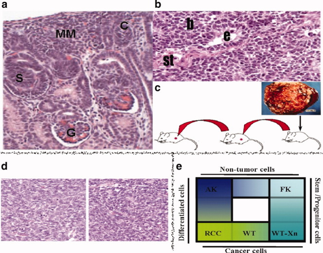
Strategy for the identification of human renal stem/progenitor markers. (A): Histological appearance of normal fetal kidney. (B): Histological appearance of primary WT. WT arises from multipotent renal embryonic precursors that undergo partial differentiation arrest, leading to a tri-phasic appearance of undifferentiated blastema (b) that resembles the MM, as well as differentiated tubular epithelial (e), and stromal (st) elements. (C): Establishment of WT-xenografts (Xn). Primary WT were implanted into SCID mice and then serially propagated, eventually leading to enrichment of stem/progenitor cells (blastema) at the expense of differentiated elements (seen in [D]). (E): Renal “stemness” markers are those elevated in microarrays of both stem-like WT-xenografts and human fetal kidneys, but not renal cell carcinoma or adult kidneys. Abbreviations: AK, adult kidney; C, C-shaped body; FK, fetal kidney; G, glomerulus; MM, metanephric mesenchyme; RCC, renal cell carcinoma; S, S-shaped body; WT, Wilms' tumor.