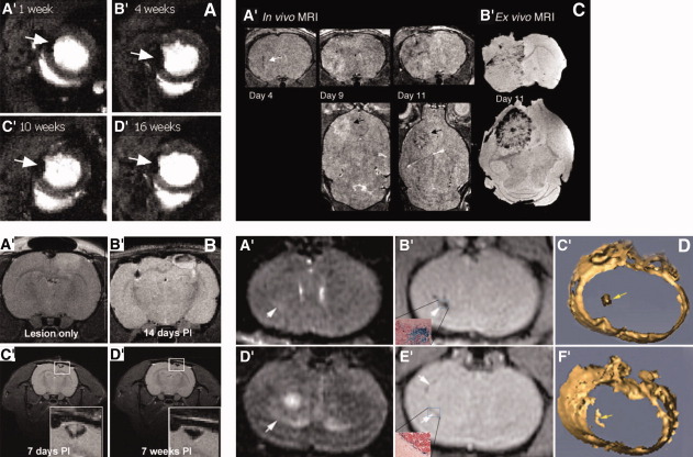
Figure 2.
MRI of superparamagnetic iron-oxide nanoparticles (SPION)-labeled stem cells showing their persistence, migration, and tumor homing in vivo. (A): Demonstrates long-term mesenchymal stem cell (MSC) traceability using SPION. Rat MSCs labeled with iron particles injected into the infracted heart could be detected as hypointense regions from 1 week and detected for up to 16 weeks. Volume of the signal void reduced to lesser extent in severely infarcted hearts in comparison with milder infarcts. ©AlphaMedPress, April 20th, 2006; Reprinted from [134], with permission from Wiley-Liss, Inc. a subsidiary of John Wiley & Sons, Inc. (B): Migration of MSCs to site of pathology: Endoderm (SPION)-labeled rat MSCs migrate toward the lesion of brain (A′) could be detected by MR up to 7 weeks after implantation (C′ and D′) in the contralateral hemisphere MSCs (B′). Reprinted from [7], with permission from Macmillan Publishers Ltd., © 2007 Nature Publishing Group. (C): Tumor homing and engraftment by Sca-1 positive bone marrow (BM) cells (target the tumor vasculature) by MRI: Serial MRI in tumor bearing mice that received magnetically labeled Sca-1+ BM cells. (A′): Three-dimensional (3D) RARE images show dark regions developing within and around tumors due to incorporation of labeled cells into the vasculature and parenchyma of tumor, images are acquired on day 4, 9, and 11. By day 11, a dark rim appear on the tumor periphery. (B′): Corresponding ex vivo gradient images of the same mouse on day 11. MR evidence of labeled cell incorporation demonstrates that neovascularization occurs primarily at the tumor periphery in the later stages of tumor development. Reproduced from [135], with permission from (This research was originally published in Blood) © 2007 The American Society of Hematology. (D): Tumor homing by SPION-labeled MSCs: The pattern of MSCs distribution, their incorporation and migration could be tracked using 1.5-T MR imaging following i.v. injection of SPION/green fluorescent protein-labeled cells. MSCs distribution throughout the tumor on day 7 (B′) was shown by a well-defined dark hypointense region. After 14 days, most MSCs were found at the tumor border (hypointense region in [D′]), (C′, F′). 3D reconstructions show the SPION-labeled MSCs as yellow structures indicated by the yellow arrows. This study demonstrates that systemically transplanted MSCs migrated toward glioma with high specificity in a temporal–spatial pattern. Reproduced from [49], with permission from ©1944-2009 by the American Association of Neurosurgeons. Abbreviations: MRI. magnetic resonance imaging.

