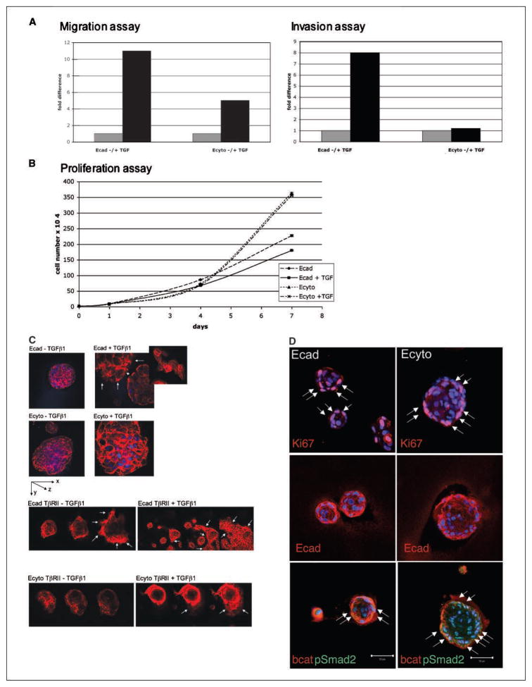
Figure 4.
Three-dimensional cultures of Ecad and Ecyto cells (spheroids) display differences in overall architecture and response to TGFβ1. A, TGFβ1 stimulation results in increased migration and invasion in both Ecad and Ecyto cells. Gray column, without TGFβ1 stimulation; black column, with TGFβ1 stimulation. B, proliferation assays show growth-inhibitory effects of TGFβ1 in Ecad cells but no changes in proliferation of Ecyto cells. C, Ecad spheroids are smaller compared with Ecyto spheroids, reflecting differences in cell-cell adhesion. TGFβ1 stimulation results in disruption of the integrity of the Ecad spheroids (arrows, cells in monolayer after TGFβ1 stimulation), which is not evident in Ecyto spheroids. Inset, cells in monolayer after TGFβ1 stimulation as well as stress fiber formation. White arrows, Ecad-TβRII spheroids disperse even in the absence of TGFβ1 and form monolayers to a greater extent in the presence of TGFβ1. This response is partially restored but delayed in Ecyto-TβRII spheroids in contrast to Ecyto spheroids that are resistant to the effects of TGFβ1. Spheroids are stained with TRITC-conjugated phalloidin. D, immunofluorescence of Ecad and Ecyto spheroids with antibody against Ki-67 (white arrows) shows fewer positive cells in the smaller Ecad spheroids. E-cadherin antibody shows localization of endogenous E-cadherin to the cell membrane in Ecad spheroids but only punctates cytoplasmic staining in Ecyto spheroids. White arrows, pSmad2 is localized to the nuclei of both Ecad and Ecyto spheroids. Bar, 50 μm.

