Figure 3.
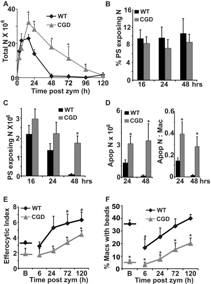
Exaggerated and prolonged neutrophilia characterizing zymosan peritonitis in CGD mice is accompanied by impaired in vivo efferocytosis by macrophages. Peritonitis was induced as in Figure 1 and peritoneal cells analyzed at the times indicated (see “Methods”). (A) Total neutrophils [N] were counted. (B) Percent of neutrophils exposing PS (annexin V–positive staining), and (C) absolute numbers of PS exposing neutrophils were determined. (D) Apoptotic neutrophils identified by nuclear morphology and the ratio of apoptotic neutrophils to macrophages were determined microscopically. (E) Efferocytic Index was determined by microscopic examination of macrophages lavaged from the inflamed peritonea. (F) Carboxylated beads were injected into the peritonea to measure in vivo efferocytic capability, and 1 hour later, the percentage of lavaged peritoneal macrophages positive for beads was determined by flow cytometry. [B] indicates baseline without zymosan. Data represent mean ± SE; N = 8 mice per time point. *P < .02 compared with WT mice at the respective time points. #P < .02 compared with WT mice at baseline.

