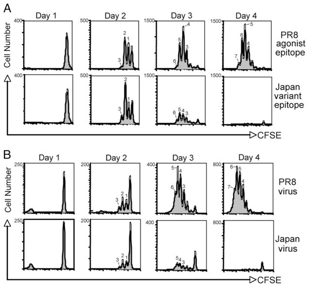
FIGURE 4.
CD8+ CL-4 T cells proliferate comparably in response to agonist and variant epitope stimulation. CFSE-labeled CD8+ CL-4 T cells were stimulated in vitro with 10−6 M PR/8 HA533–541 or Japan HA529–537 peptide-pulsed splenocytes (A) or with A/PR/8/34 or A/Japan/305/57 influenza virus-infected splenocytes (B). Division was analyzed by flow cytometry and CFSE dilution at the indicated times poststimulation. Histograms represent CD8+ cells within the live lymphocyte gate.

