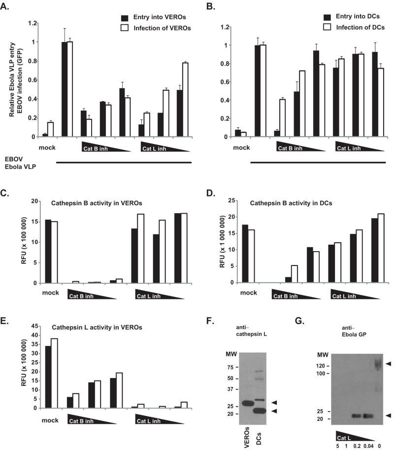
Figure 4. Effect of cathepsin B and L inhibitors on Ebola VLP entry and EBOV infection of VEROs and DCs.
VERO (A, C, E) and DCs (B, D) were pretreated with mock and decreasing concentrations of cathepsin B and cathepsin L, respectively. Also, VERO (A) and DCs (B) were mock and VLP treated or EBOVGFP-infected (denoted by bar under graphs). Solid bars represent relative percentage of VEROs (A) and DCs (B) infected by VLPs and white bars represent relative percentage of VEROs (A) and DCs (B) infected by EBOVGFP. Cathepsin B (C, D) and cathepsin L (E) activity was measured in duplicate (solid and white bars) using a fluorogenic substrate and expressed as relative fluorenscence units (RFU) using equivalent amounts of pretreatred VEROs (C, E) and DCs (D). (F) Equivalent amounts of VERO (lane 1) and DC (lane 2) lysates (same lysates used to determine cathepsin activity) were subjected to SDS-PAGE and western blotting using anti-cathepsin L antibody (204106, R&D systems, Minneapolis, MN). Arrow denotes positions of Cathepsin L species. (G) VP40+GP VLPs were incubated for 1.5 hours with decreasing concentrations of recombinant cathepsin L (5, 1, 0.2, 0.04 μg/mL) and subjected to western blotting using a polyclonal anti-GP antibody. Arrow denotes positions of GP species.

