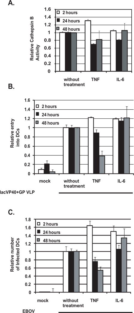
Figure 5. Effect of TNFα and IL-6 treatment on DC cathepsin B activity and susceptibility of DCs to EBOV entry and infection.
DCs were pretreated for 2 hours, 24 hours and 48 hours (A, B, C) with mock, TNFα and IL-6 cytokines. (A) DC cell lysate was then tested for total cathepsin B activity and is represented as relative cathepsin B activity. This experiment was repeated with similar results. (B and C) Pretreated DCs were subjected to VLP (lacVP40+GP) entry assay (B) and EBOVGFP infection (C). Shown is the relative entry and infection if the mock control is set to 1. This experiment has been repeated with similar results with at least six different donors.

