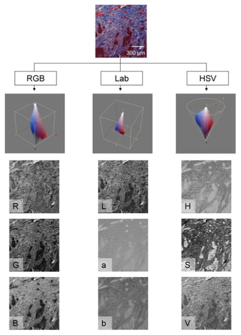
FIGURE 2.

A small histology sample, immunohistochemically stained with the pan-CK with Masson's Trichrome (Mod 2) counterstain, is shown along with the corresponding represented in the Red–Green–Blue (RGB), CIE L*a*b* (Lab), and Hue-Saturation-Value (HSV) color spaces.
