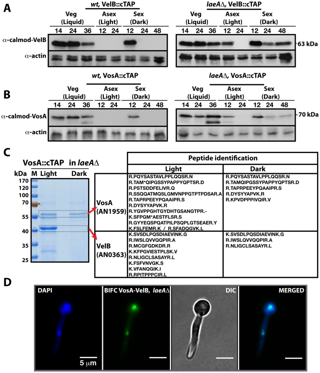
Figure 3. LaeA control of VosA and VelB protein levels and the VosA-VelB complex formation.
(A) VelB::cTAP and (B) VosA::cTAP fusion protein levels detected by α-calmodulin antibody during different developmental stages in wild type (wt) and laeAΔ strains at 37°C. α-actin served as internal control. Protein crude extracts (80 µg) were loaded in each lane. (C) Brilliant blue G-stained 10% SDS-polyacrylamid gel of VosA::cTAP and identified polypeptides (Table S5) in laeAΔ strain grown in the light and dark are given. (D) BIFC interaction of the nuclear VosA-VelB complex in laeAΔ strain. N-EYFP::VosA interacts with C-EYFP::VelB. Nuclei were counterstained with DAPI (blue).

