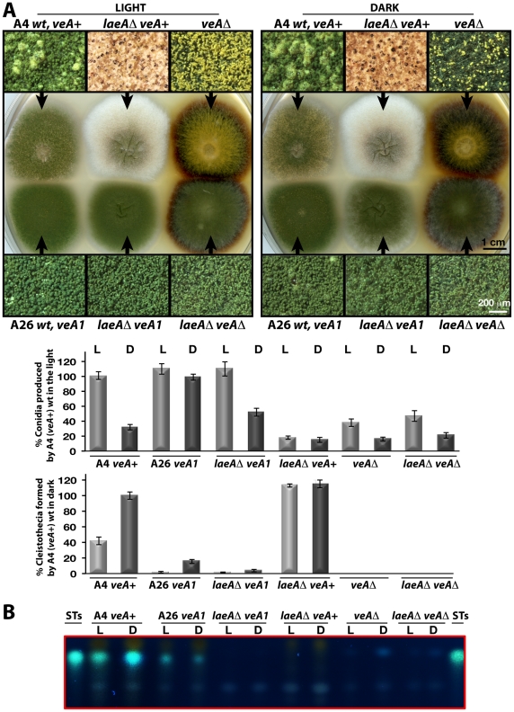
Figure 5. LaeA-VeA as regulators of development and secondary metabolism.
(A) Colony morphologies, quantifications of asexual spore (conidia, in light) and fruiting body (cleistothecia, in dark) formations of (A4) veA+, (A26) veA1, laeAΔ/veA+, laeAΔ/veA1, veAΔ, laeAΔ/veAΔ strains grown on the plates at 37°C for 5 days in the light asexually or in the dark sexually. For the quantification of conidia or cleistothecia, the 5×10 mm2 sectors from 5 independent plates were used and the standard deviations are indicated as vertical bars. veA+ strains conidiation and cleistothecia levels were used as standard (100%). (B) The secondary metabolite sterigmatocystin (ST) production levels of the strains from (A) examined by TLC. 5×10³ conidia were point-inoculated at the center of the plates that were kept either in white light (90 µWm2) or in dark.

