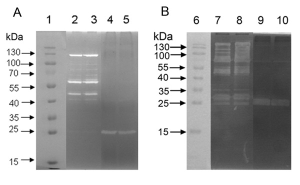
Figure 5.
Zymographic analysis of S. epidermidis 1457ΔlytSR. Extracellular and cell surface proteins were isolated, and 30 μg of each was separated in SDS-polyacrylamide gel electrophoresis gels containing 2.0 mg of M. luteus (A) or S. epidermidis (B) cells/ml. Murein hydrolase activity was detected by incubation overnight at 37 °C in a buffer containing Triton X-100, followed by staining with methylene blue. Lanes: 1 and 6, molecular mass marker; 2 and 7, cell wall protein from 1457ΔlytSR strain; 3 and 8, cell wall protein from wild type strain; 4 and 9, extracellular protein from 1457ΔlytSR strain; 5 and 10, extracellular protein from wild type strain. The results are representative of three independent experiments.

