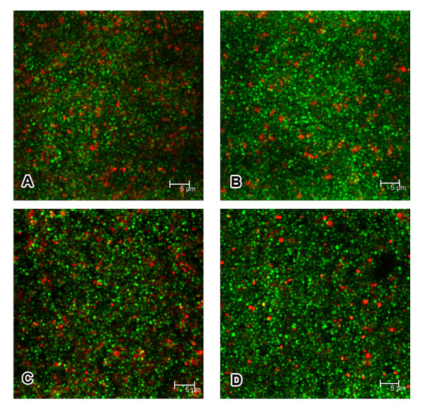
Figure 8.
Confocal photomicrographs of 24-hour-old biofilms. Biofilms containing S. epidermidis 1457 strains wild-type (A), ΔlytSR (B), ΔlytSR(pNS-lytSR) (C) and ΔlytSR(pNS) (D) were visualized by using the live/dead viability stain (SYTO9/PI). Green fluorescent cells are viable, whereas red fluorescent cells have a compromised cell membrane, as indicative of dead cells. Scale bars = 5 μm. The result is a stack of images at approximately 0.3 μm depth increments and represents one of the three experiments.

