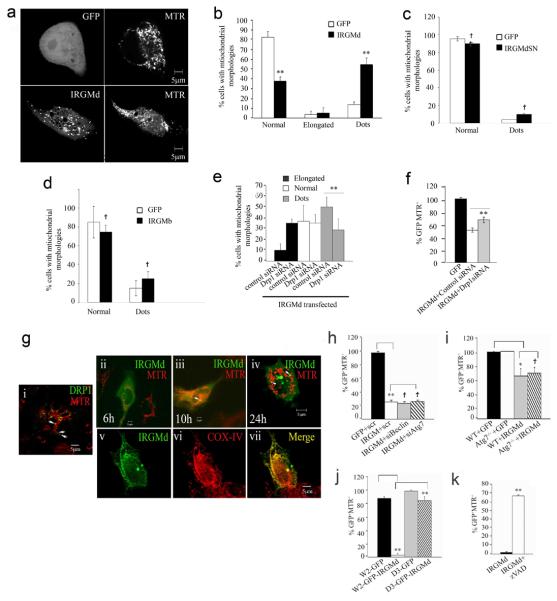
Fig. 6. IRGMd translocates to mitochondria, induces mitochondrial fragmentation and causes loss of mitochondrial Δψm independent of autophagic but dependent on apoptotic machinery.
a. HeLa cells transfected with GFP or GFP-IRGMd for 24 h were stained with MTR and imaged. b-d. Quantification of mitochondrial morphologies in cells transfected with IRGMd, IRGMdS47N, or IRGMb fusions. e,f. IRGM acts in concert with DRP1. Cells were transfected with siRNAs (48 h) and GFP-IRGMd, and mitochondrial morphologies (e; 24 h) or MTR staining (f; % of GFP+ cells that were also MTR+ at 48 h) quantified. g. IRGMd translocates (ii-iv) from the cytosol to mitochondria and colocalizes with COX IV (v-vii). Cytosol-to-mitochondria translocation and mitochondrial localization was compared with the typical appearance of steady state Drp1 distribution (i). h. HeLa cells, co-transfected with GFP-IRGMd and control, BECN1 (Beclin 1) or ATG7 siRNA, were labeled with MTR 48 h post-transfection, imaged by live microscopy, and % of GFP+ cells that were MTR+ quantified. i. Atg7 wild type (WT) or Atg7−/− MEF transfected with GFP-IRGMd for 48 h were analyzed for mitochondrial staining as in g. j. Wild type W2 (Bax/Bak+/+) or mutant D3 (Bax/Bak−/−) BMK cells were transfected with GFP-IRGMd for 48 h and % of GFP+ cells that were MTR+ quantified. k. HeLa cells were transfected with GFP-IRGMd, treated with z-VAD, and after 48 h stained with MTR. Data, means ± SEM (n=3). †P≥0.05,*P<0.05, **P<0.01, (t-test).

