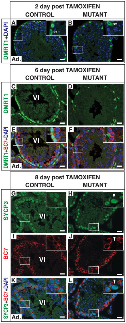
Figure 4. Kinetics of the switch from mitosis to meiosis in mutant spermatogonia.
Conditional deletion of DMRT1 in adult testes using the tamoxifen inducible cre transgene Ubc-cre/ERT2. (A,B) Section IF for DMRT1two days after tamoxifen injection showing DMRT1 in spermatogonia and Sertoli cells (SC) in control but only in Sertoli cells in mutant. (C-F) Section IF six days after tamoxifen injection. BC7-positive cells at stage VI are primary spermatocytes (late zygotene to mid-pachytene) with underlying layer of DMRT1-positive B spermatogonia (SC: Sertoli cell). Inset shows missing layer of spermatogonial cells in mutant; DMRT1-positive cells in mutant are Sertoli cells. (G-L) Section IF eight days after tamoxifen injection. In stage VI control tubule the germ cells underlying the layer of BC7- and SYCP3-positive primary spermatocytes are negative for SYCP3. In mutant tubules with abundant BC7- and SYCP3-positive primary spermatocytes, underlying cells have SYCP3 distribution typical of leptotene spermatocytes (arrowheads), not normally found together with BC7-positive spermatocytes. Scale bars: 20 microns.

