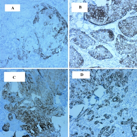
Fig. 2.
K19 immunostain in OSCC groups. Objective lens magnification of ×10, and resolution of 1.10 μm. a Well-differentiated OSCC with abundant keratin pearl formation. Note the pale staining of the outer layer of the invasive epithelial islands. b Well-differentiated OSCC with rare keratin pearl formation. Note the diffuse staining of invasive islands. c Moderately differentiated OSCC showing diffuse staining of invasive islands. d Poorly differentiated OSCC with diffuse positivity

