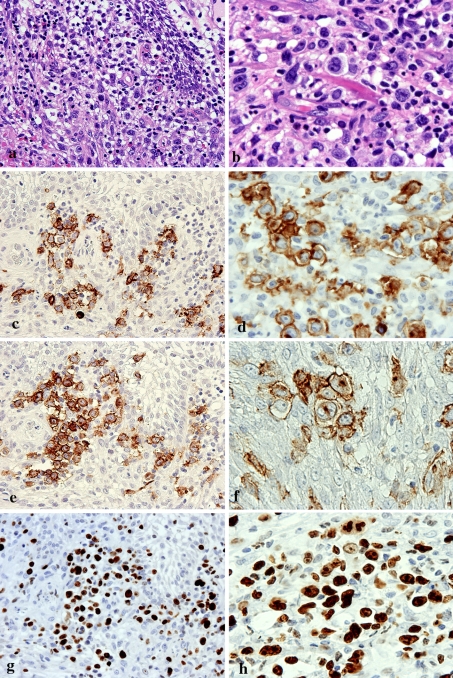
Fig. 4.
Immunohistochemical features of the re-biopsy specimen. a, c, e, g, showing an edematous submcosal stroma (original magnification × 400). b, d, f, h Granuloma formation (original magnification × 400). a HE staining of the edematous submcosal stroma. b HE staining of the granulomatous area. c, d Atypical large cells show positivity for CD20. e, f An atypical large cell positive for CD30. f Reed-Sternberg (R-S)-like cells show positivity for CD30. g, h Similarity, nuclei of large round atypical cells are markedly positive for Ki-67 (labeling index exceeding 60–70%)

