
Figure 2.
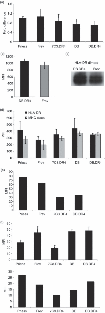
Comparable expression of MHC class II messenger RNA and protein in Danon and wild-type B-LCL. (a) Total RNA was extracted from various wild-type or Danon B-LCL, complementary DNA was synthesized, and quantitative reverse transcription–polymerase chain reaction analysis performed using primers specific for HLA-DRα chain or for GAPDH as a control. Data are representative of the fold difference in messenger RNA expression observed in three independent experiments. (b) Wild-type Frev or LAMP-2-deficient DB.DR4 cells were incubated with L243 antibody to detect total surface HLA-DR and then stained with a fluorescein isothiocyanate-conjugated F(ab′)2 fragment of goat anti-mouse IgG secondary antibody. The mean fluorescence intensity (MFI) as measured by flow cytometry indicates the level of surface HLA-DR, and data are the average MFI of three independent experiments. (c) Cell lysates were prepared from LAMP-2-deficient DB.DR4 or wild-type Frev B-LCL, the proteins resolved by gel electrophoresis under non-reducing conditions to preserve MHC class II dimers, and immunoblotted with an antibody to HLA-DRα chain. Data are representative of at least five independent experiments. The ratios of HLA-DRαβ dimers to GAPDH as a loading control were 1·5 and 1·3 for DB.DR4 and Frev, respectively. (d) Various wild-type or Danon B-LCL were first permeabilized and then incubated with L243 or W6/32 antibody to detect total intracellular HLA-DR or MHC class I molecules, respectively. Cells were then stained with a phycoerythrin-conjugated F(ab′)2 fragment of rabbit anti-mouse immunoglobulin secondary antibody. The MFI as measured by flow cytometry indicates the levels of total surface or intracellular MHC class I or class II molecules. Data are the average MFI of three independent experiments. (e) Wild-type Priess and Frev expressing endogenous HLA-DR4 and wild-type 7C3.DR4 or LAMP-2-deficient DB.DR4 transfectants were incubated with 3.5.9-13F10 antibody to detect surface HLA-DR4β chains and then stained with a Cy2-conjugated F(ab′)2 fragment of donkey anti-rat IgG secondary antibody. The MFI as measured by flow cytometry indicates the level of surface HLA-DR4, and data are representative of more than three independent experiments. (f) Various wild-type or Danon B-LCL were first permeabilized and then incubated with MaP.DM1 (top panel) or a monoclonal antibody to HLA-DO (bottom panel) to detect total intracellular HLA-DM or HLA-DO molecules, respectively. Cells were then stained with a phycoerythrin-conjugated F(ab′)2 fragment of rabbit anti-mouse immunoglobulin secondary antibody. The MFI as measured by flow cytometry indicates the levels of total intracellular HLA-DM or HLA-DO molecules. Data for HLA-DM staining are the average MFI of three independent experiments while the data for HLA-DO staining are a representative experiment.