
Figure 5.
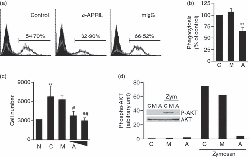
The stimulation of APRIL inhibited phagocytosis of opsonized zymosan and transmigration in THP-1 cells through suppressing the activities of phosphatidyl inositol 3-kinase (PI3K). (a) THP-1 cells were pre-treated for 30 min with 10 μg/ml of anti-APRIL monoclonal antibody (mAb) or mouse immunoglobulin G (mIgG). Cells were then incubated with 30 μg/ml of fluorescence-labelled opsonized zymosan. Three hours later, fluorescence levels in each sample were measured using flow cytometry. (b) The assay in (a) was repeated three times and the flow cytometry results were subjected to a statistical analysis after setting the control measurement as 100%. Values indicate mean + SEM. **P < 0·01, when compared with the control. C, no treatment control; M, mIgG; A, anti-APRIL. (c) Transmigration potential of THP-1 cells pre-treated with 20 μg/ml of mIgG (M) or 10 and 20 μg/ml of anti-APRIL mAb (A) was measured with 10 μg/ml of fibronectin as a chemoattractant. Experiments were repeated three times for statistical analysis. Numbers represent mean + SEM of migrated cell numbers. **P < 0·01, when compared with the no treatment control (N). #P < 0·05, ##P < 0·01, when compared with the control (C). (d) THP-1 cells were stimulated with 30 μg/ml of opsonized zymosan in the presence or absence of 10 μg/ml of anti-APRIL mAb (A) or mouse IgG (M) for 3 hr. Cell lysates were obtained after 2 hr for the Western blot analysis of AKT and phospho-AKT (inset). Phospho-AKT band intensities were then measured with densitometer and normalized with band intensities of corresponding AKT. Results are representative for three independent experiments.