
Figure 5.
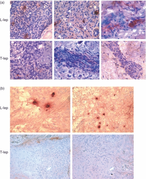
Immunoglobulin M (IgM) and IgA expression in leprosy lesions (T-lep, tuberculoid leprosy lesions; L-lep, lepromatous leprosy lesions). Representative sections from skin biopsy specimens of tuberculoid and lepromatous lesions stained by the immunoperoxidase method with monoclonal antibodies specific for IgM (a) and IgA (b). Multiple IgM- and IgA-positive cells were identified in the dermis of L-lep patients. In contrast, IgM- and IgA-positive cells were infrequent in the T-lep patients. Original magnification: × 400.