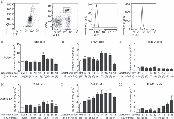
Figure 1.

Increased proliferation of CD8 T cells during normal pregnancy. Pregnant and unmated (UM) C57BL/6 mice were injected with bromodeoxyuridine (BrdU) for the 24 hr before death, and parallel samples from each mouse were analysed for proliferation by BrdU incorporation, and apoptosis by the terminal deoxynucleotidyl-transferase (TdT)-mediated dUTP nick-end labelling (TUNEL) assay. (a) Representative analysis of flow cytometric data from a day 12 pregnant mouse. Left to right: forward versus side scatter plot of whole spleen, gating of CD8+ TCR-β+ cells, histogram analysis for the proportion of BrdU+ cells, and histogram analysis for the proportion of TUNEL+ cells. (b) Numbers of CD8 T cells in the spleen were calculated by multiplying the total number of cells counted by the proportion of CD8+ TCR-β+ cells obtained by flow cytometry. (c) Number of splenic CD8+ T cells that were BrdU+ on each gestational day. (d) Number of splenic CD8+ T cells positive for TUNEL staining throughout pregnancy. (e) Numbers of CD8 T cells in the uterine draining lymph nodes were calculated by multiplying the total number of cells counted by the proportion of CD8+ TCR-β+ cells obtained by flow cytometry. (f) Number of uterine draining lymph node CD8+ T cells that were BrdU+ on each gestational day. (g) Number of uterine lymph node CD8+ T cells positive for TUNEL staining throughout pregnancy. In each graph symbols and error bars depict mean and standard error of the mean (SEM). Numbers in parenthesis represent the total number of mice analysed at each time-point. Statistical significance was determined by one-way analysis of variance with Dunnett's post-test using UM controls for comparison *P < 0·05, **P < 0·01.
