
Figure 1.
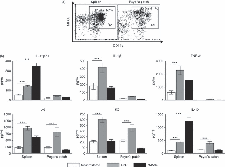
Peyer's patch dendritic cells (PP DCs) secrete a limited profile of cytokines compared with spleen DCs. (a) Spleen and PP DCs were isolated by autoMACS selection of CD11c+ cells. The purity of cells is demonstrated. Values in the upper right indicate mean ± SEM percentage of cells falling in the gate, n = 5. (b) Following CD11c+ isolation cells were cultured for 18 hr with lipopolysaccharide (LPS; 1 μg/ml) or phorbol 12-myristate 13-acetate/ionomycin (PMA/Io; 5 ng/ml PMA, 1 μg/ml Io) or left unstimulated. Supernatants were collected and interleukin-12 (IL-12) p70, IL-1β, tumour necrosis factor-α (TNF-α), IL-6, IL-10 and keratinocyte chemoattractant (KC) levels were determined by electrochemical enzyme-linked immunosorbent assay. Bars represent mean ± SEM. Significance determined by one-way analysis of variance, Tukey post-hoc test, ***P < 0·001, n = 3 to n = 9.