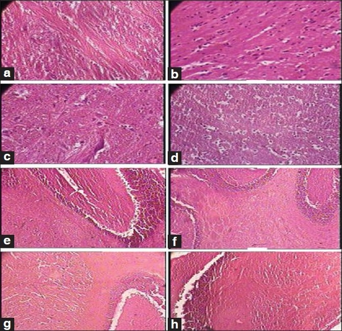
Figure 1.

Photomicrographs of sections of hindbrain and cerebellum Note the normal cytoarchitecture in all the groups, (a): Hindbrain of the control group (1 × 400 magnification), (b, c): Hindbrain of the therapeutically equivalent dose group (1 × 400 magnification), (d): Hindbrain of the therapeutically equivalent dose × 10 group (1 × 400 magnification), (e): Cerebellum of the control group (1 × 100 magnification), (f): Cerebellum of the therapeutically equivalent dose group (1 × 100 magnification), (g): Cerebellum of the therapeutically equivalent dose × 5 group (1 × 100 magnification), (h): Cerebellum of the therapeutically equivalent dose × 10 group (1 × 100 magnification)
