
Fig. 2.
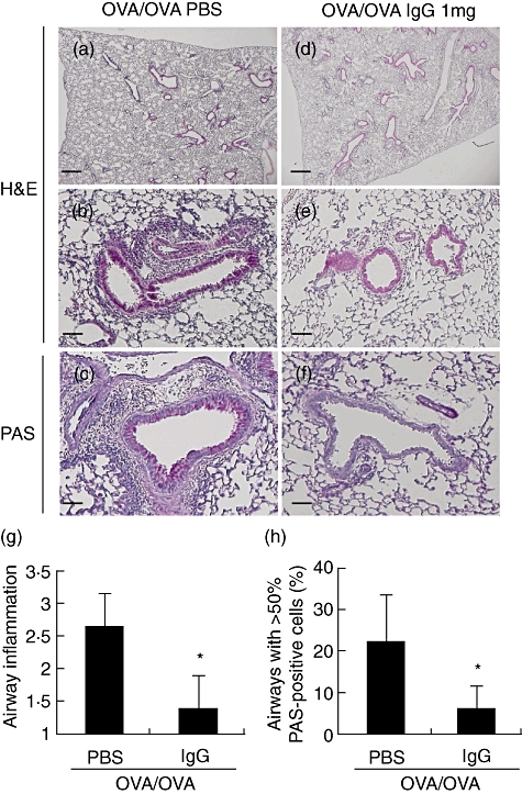
Effects of intravenous (i.v.) immunoglobulin G (IVIgG) on the development of airway inflammation and mucus production. Paraffin-embedded sections of lung of sensitized and challenged mice were prepared. Representative histological findings for lungs stained with haematoxylin and eosin (H&E) or periodic-acid Schiff (PAS) are shown. (a–c) Lung tissue from challenged mice [ovalbumin (OVA)/OVA + phosphate-buffered saline (PBS)]. (d–f) Lung tissue from mice administered with 1 mg of IgG (OVA/OVA + IgG). (a,d) Low-power field of the H&E-stained sample. (b,e) High-power field of the H&E-stained sample. (c,f) High-power fields of the PAS-stained sample, which shows mucus stained red by PAS. Scale bar equals 100 µm on low-power fields (a,d) and 50 µm on high-power fields (b,c,e,f). (g) Scores for peribronchial and perivascular inflammation are shown. (h) The number of airways free of mucus-producing cells or containing > 50% PAS-positive-cells are shown. *Significant differences (P < 0·05) versus PBS.