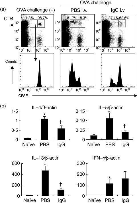
Fig. 4.

Effects of intravenous (i.v.) immunoglobulin G (IVIgG) on antigen presentation and T helper type 2 (Th2) differentiation. (a) Flow cytometric analysis of antigen presentation to ovalbumin (OVA)-specific CD4+ T cells in vivo is shown. Carboxyfluorescein succinimidyl ester (CFSE)-labelled CD4+ OVA-specific OTII lymphocytes (5 × 10⁶ cells) were transferred into mice challenged with OVA and administered with rabbit IgG or phosphate-buffered saline (PBS). At 24 h after the last challenge, the proliferation of CD4+ T cells in thoracic lymph nodes (TLNs) was analysed. The histogram shows the reduction of CFSE intensity, which indicates cell division and proliferation of the transferred CFSE+ CD4+ cells. One representative experiment of three with similar results is presented. In the inset boxes the percentages of divided (left box) or undivided cells (right box) among CFSE+ CD4+ cells subsets in representative data are shown. (a) The mRNA levels of Th2 cytokines [interleukin (IL)-4, IL-5, IL-13] and Th1 cytokine [interferon (IFN)-γ] in co-culture of OTII CD4+ T cells and lung CD11c+ cells with OVA323–339 peptide are shown. CD4+ cells (2·5 × 105 cells) isolated from spleens of OTII mice using CD4-microbeads and magnetic-activated cell sorting (MACS) system were stimulated with the OVA peptide (5 µg/ml) and lung CD11c+ cells (2·5 × 104 cells) from IgG/PBS-administered mice. After 6 h, mRNA levels of cytokines were evaluated by real-time polymerase chain reaction (PCR) *Significant differences (P < 0·05) versus naive mice. †Significant differences (P < 0·05) versus PBS.
