Fig. 1.

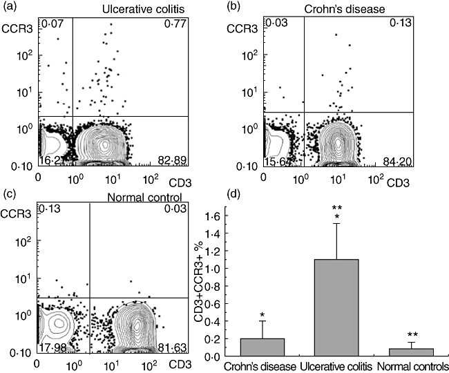
Peripheral T cells from ulcerative colitis (UC) patients are enriched preferentially in CCR3+ cells. The presence of CCR3 was assessed by flow cytometry in peripheral T lymphocytes from ulcerative colitis (UC, n = 20), Crohn's disease (CD, n = 22) and normal controls (NCs, n = 20), isolated from peripheral blood mononuclear cells (PBMCs) via magnetic separation based on CD3 expression. (a–c) Representative fluorescence activated cell sorter (FACS) images (CD3+CCR3+) from patients with UC, CD and NCs. (d) Each column represents the mean value ± standard error of the mean of CD3+ CCR3+ cells of different groups derived from flow cytometry (*P < 0·05 between UC and CD patients and **P < 0·05 between patients with UC and NCs).
