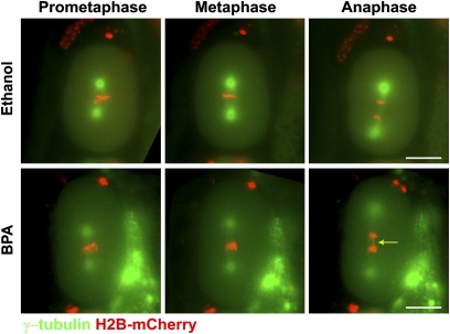
Fig. 4.
BPA exposure results in chromosome segregation defects during early embryonic cell division. Time-lapse analysis is shown of the first embryonic division in ethanol- and BPA-exposed H2B::mCherry; γ-tubulin::GFP embryos (n = 60 per condition). Chromosomes (red) fail to properly align along the metaphase plate in the BPA-exposed embryo. The arrow indicates a chromatin bridge observed in the same embryo during anaphase. (Scale bars, 20 μm.)

