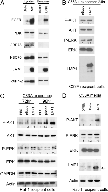
Fig. 3.
Exosomes and conditioned media from LMP1-expressing C33A cells activate ERK and AKT in recipient cells. (A) Cell lysates and purified exosomes from C33A-LMP1 or C33A-pBabe cells were analyzed by immunoblotting with antibodies against EGFR, p85, GRP78, HSC70, LMP1, flotillin-2, actin, and S12 mAb for LMP1. (B) Untransfected C33A cells were incubated with C33A-pBabe or LMP1 exosomes for 24 h in serum-free media, and the indicated proteins were identified by immunoblotting of cell lysates. (C) C33A exosomes were incubated with Rat-1 cells for 72 or 96 h, and cell lysates were analyzed for the indicated proteins by immunoblotting. Numbers indicate fold change over PBS control. (D) Media alone (DMEM) or conditioned media from C33A-pBabe or C33A-LMP1 was clarified and added to Rat-1 cells every 24 h for 5 d. pAKT and pERK intensities in all experiments were normalized to total protein levels, and the relative values to the control are indicated in each channel. Representative blots from two to four independent experiments are shown.

