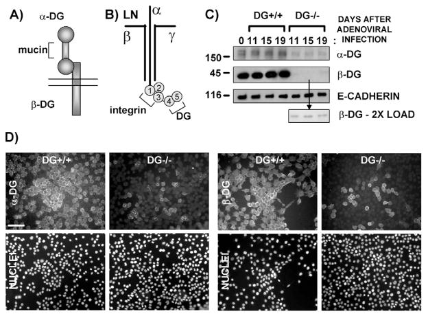
Fig. 1.
Generation of DG+/+ and partial-DG−/− MEC populations by adenoviral infection of immortalized mouse MECs. (A) Diagram of DG, including the extracellular α-DG subunit, with central mucin domain, and the transmembrane β-DG subunit. (B) Diagram of laminin-111 (LN), including the three subunits (α, β, γ), and the five C-terminal LG domains, with respective receptor binding sites. (C) Western blot of cell extracts (5 μg protein) prepared on different days after infection of the immortalized, floxed DG mouse MEpG cell line with control or Cre-recombinase-expressing adenovirus to generate DG+/+ and partial-DG−/− cell populations, respectively. The first lane (far left) represents uninfected cells at time 0. Blots were incubated with antibodies specific for α-DG, C-terminal β-DG or E-cadherin (loading control), followed by HRP-conjugated secondary antibodies. Sizes of molecular mass markers are shown in kDa. (D) Vertically paired immunofluorescent images of DG+/+ and partial-DG−/− MEpG cell populations using primary antibodies specific for α-DG or C-terminal β-DG, followed by FITC-labeled secondary antibodies (upper panel). Nuclei were stained with propidium iodide (bottom panel). Bar, 60 μm.

