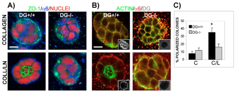
Fig. 2.

Loss of polarity in DG−/− colonies grown in a 3D matrix of collagen I–laminin-111. DG+/+ and DG−/− MEpG cells were grown in a 3D matrix of collagen I or collagen I–laminin-111 and co-immunostained. Confocal immunofluorescent images were taken at colony centers. Bars, 10 μm. (A) Staining using anti-ZO-1 and anti-α6 integrin antibodies, visualized with FITC- (green) and Cy5- (blue) labeled secondary antibodies, respectively, and propidium iodide to stain nuclei (red). (B) Staining using antibodies against α6 integrin and C-terminal β-DG (insets), detected with Rhodamine- (red) and Cy5- (blue changed to white for easier visualization) labeled secondary antibodies, respectively. Actin was seen using Alexa Fluor-488–phalloidin (green). Overlap between actin and α6 integrin staining appeared yellow. (C) Quantification of polarity in DG+/+ and DG−/− colonies grown in collagen I (C) or collagen I–laminin-111 (C/L) using ZO-1 as a polarity marker. Results are shown as the mean ± s.e.m. of four to six independent experiments, each with triplicate or quadruplicate counts. *P<0.01, for all paired combinations.
