Fig. 3.
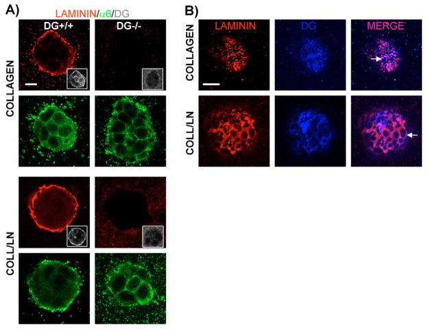
Loss of laminin binding and DG colocalization on the surface of DG−/− cells grown in a 3D matrix of collagen I or collagen-I–laminin-111. (A) Vertically paired confocal immunofluorescent images of DG+/+ and DG−/− MEpG cells grown in collagen I or collagen-I–laminin-111. Samples were co-immunostained with laminin, α6 integrin and C-terminal β-DG (insets) antibodies, followed by Rhodamine- (red), FITC- (green), and Cy5- (blue changed to white for easier visualization) labeled secondary antibodies, respectively. Images were taken at colony centers. (B) Confocal immunofluorescent images taken at the cell surface of DG+/+ colonies shown in A to reveal co-staining for laminin and β-DG, and their extent of co-localization. Arrows point to arrays of laminin. Bars, 10 μm.

