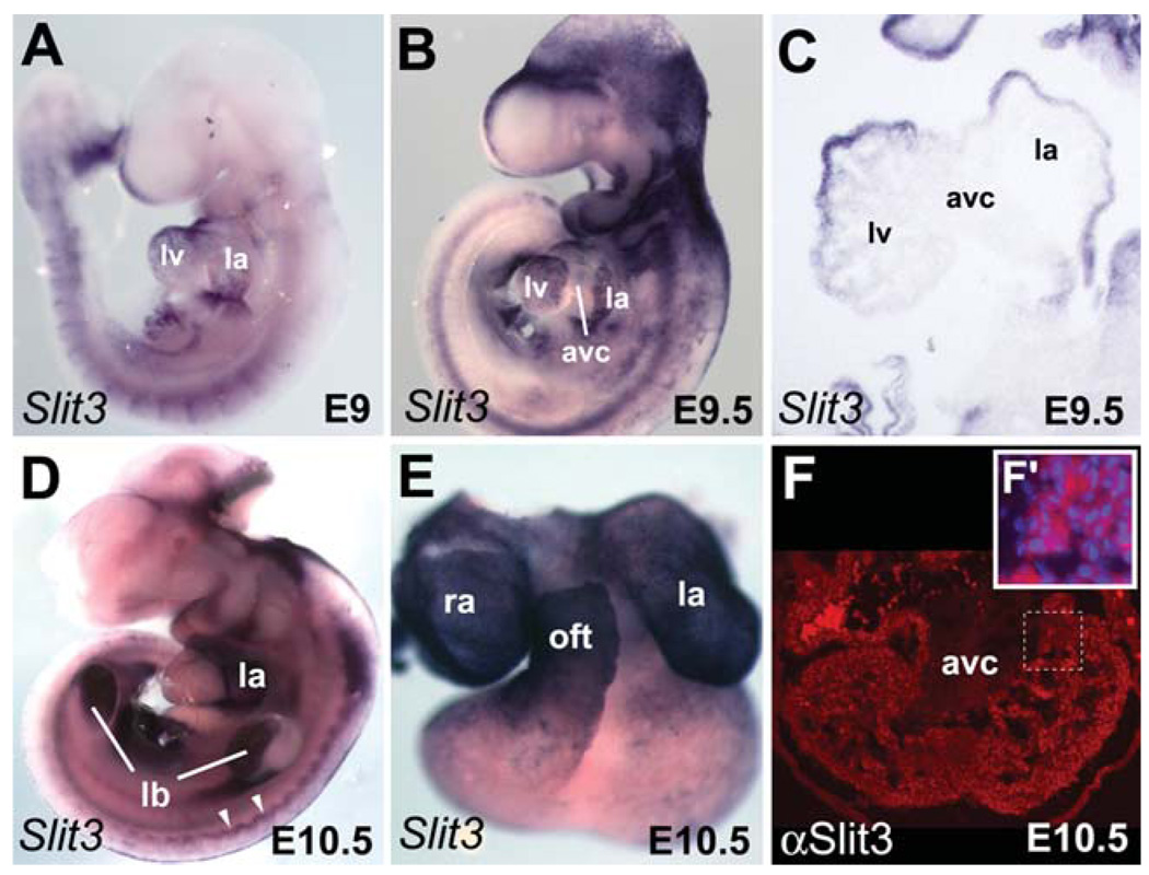
Fig. 4.
Dynamic expression pattern of Slit3 during heart development. In situ hybridization (A–E) and immunofluorescence (F) were used to detect spatial expression of Slit3 in hearts from embryonic day (E) 9 to E10.5. A,B: Expression of Slit3 is detected in the whole heart of E9 embryo, whereas it is down-regulated in the AVC of late E9.5 embryo. C: Section (same embryo as depicted in B) showing expression of Slit3 in the chambers but not in the AVC. Note weak expression of Slit3 in the trabeculae of the left ventricle. D: Lateral view of E10.5 embryo. High expression of Slit3 is detected in the left atrium. Note Slit3 expression in the limb buds and the dermomyotome (white arrowheads). E: Dissected heart from the embryo shown in D. High expression is observed in the right and left atria and the outflow tract. F: Expression of Slit3 protein (red) is detected in the ventricular myocardium and trabeculae but not in the AVC of heart at E10.5. Higher magnification of the trabeculae region delimited by the dotted line is shown in the inset. F′: Immunodetection of Slit3 (red) and DAPI (blue) staining. Note Slit3 expression in the cytoplasm. ao, aorta; avc, atrioventricular canal; la, left atrium; lb, limb bud; lv, left ventricle; oft, outflow tract; pt, pulmonary trunk; ra, right atrium; rv, right ventricle.

