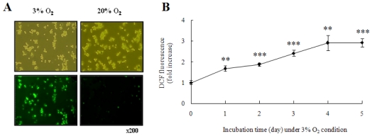
Figure 2.
Hypoxia generated intracellular ROS in THP-1 cells. (A) Representative fluorescence microscopic images showing the fluorescence of DCF oxidized by intracellular ROS in THP-1 cells grown in 20% or 3% O2 for 5 days. After cultivation, the cells were incubated with 10 μM DCFH-DA for 15 min at 37 °C and assessed by fluorescence microscopy; (B) Normalized mean fluorescence intensities (MFI) of DCF in THP-1 cells grown in 3% O2 for 1–5 days were examined by FACS analysis.

