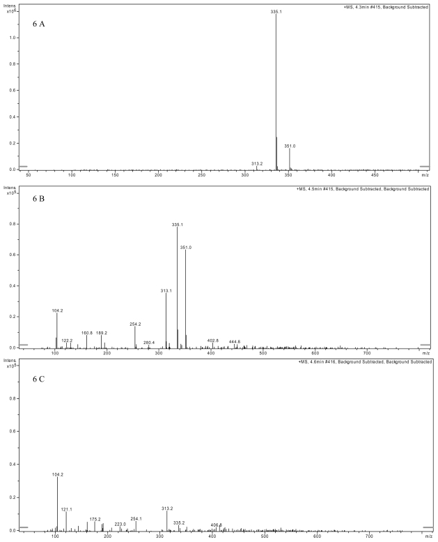
Figure 6.
Detection of AFB1 by liquid chromatography mass spectrometry. (A) LC-MS spectrum of AFB1 standard at 100 ppb showing the distinct ions: [M + H]+ at m/z = 313, [M + Na]+ at m/z = 335 and [M + K]+ at m/z = 351. (B) LC-MS spectrum of VY/2 medium supplemented with AFB1 standard at 100 ppb. The three distinct ions can clearly be distinguished. (C) LC-MS spectrum of AFB1 after 72 h treatment with M. fulvus culture supernatant. The ion at 351 disappeared while the ions at 313 and 335 were significantly reduced.

