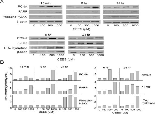
Fig. 4. Effects of CEES on expression of markers of injury and inflammation.
EpiDerm-FT™ was treated with CEES (100, 300 or 1000 μM) or control for 15 min, 6 hr, or 24 hr, as indicated. Panel A. Epidermal sheets were collected and analyzed for protein expression by Western blotting using anti-PCNA, PARP, phospho-H2AX, COX-2, 5-LOX, and LTA4 hydrolase antibodies. β-actin was used as a control to ensure equal protein loading. Panel B. Densitometry was performed on Western blots shown in Panel A. Data is presented in arbitrary units.

