
Figure 4.
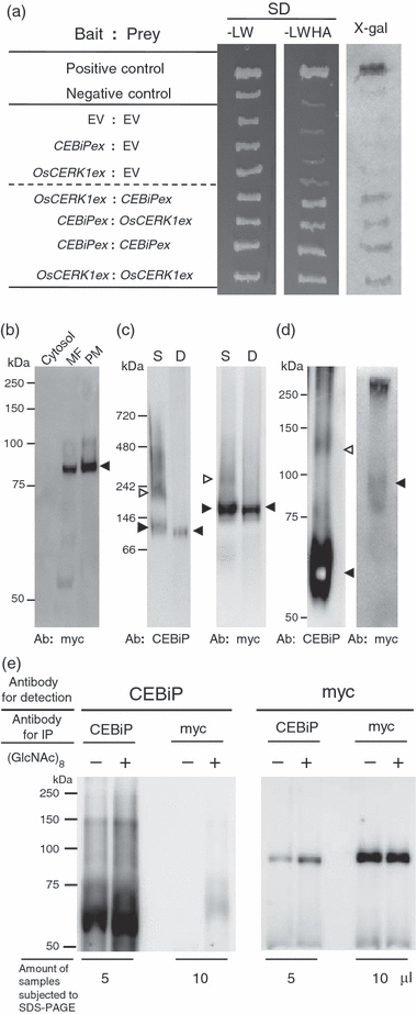
Interaction of LysM-containing receptor molecules.(a) Possible interactions between LysM-containing receptor molecules. A yeast two-hybrid assay was performed using the extracellular domains of CEBiP (CEBiPex) and OsCERK1(OsCERK1ex) as bait or prey. Growth on selective medium (SD/-LWHA) and blue colony formation in the X-gal assay indicated a positive interaction. EV, empty vector.(b) Localization of OsCERK1 in rice cells. Cytosol, plasma membrane (PM) and microsome (MF) fractions were prepared from rice cells expressing myc-tagged OsCERK1 (OsCERK1:myc), and analyzed for the presence of OsCERK1:myc by Western blotting with anti-myc antibody. The arrowhead indicates the denatured OsCERK1:myc protein.(c) Analysis of protein complex in the plasma membrane by BN-PAGE. A plasma membrane preparation from rice cells expressing OsCERK1:myc was solubilized with 0.5%n-dodecyl-β-d-maltoside and subjected to BN-PAGE. CEBiP and OsCERK1:myc were detected with anti-CEBiP antiserum (left) and anti-myc antibody (right). Microsome proteins from the same cell line were completely denatured by boiling with SDS–PAGE sample buffer and used to show the positions of the CEBiP or OsCERK1:myc monomers. S, solubilized plasma membrane proteins; D, denatured microsome proteins.(d) In vitro chemical cross-linking for the detection of protein complexes containing CEBiP or OsCERK1:myc. Plasma membranes from rice cells expressing OsCERK1:myc were treated with 3 mg/ml of DTSSP, followed by SDS–PAGE. Immunodetection was performed with anti-CEBiP antiserum (left) and anti-myc antibody (right).(e) Analysis of the CEBiP–OsCERK1 interaction by immunoprecipitation. Microsomes from rice cells expressing OsCERK1:myc pre-treated with or without (GlcNAc)8 were solubilized with 0.5% Triton X-100 and immunoprecipitated with anti-CEBiP or anti-myc antibody. The immunoprecipitates were recovered using protein A beads and eluted with SDS–PAGE sample buffer. Aliquot (5 or 10 μg) of the total eluate (60 μl) were subjected to SDS–PAGE and detected with the corresponding antibodies. The presence of CEBiP and OsCERK1:myc in the immunoprecipitates was determined using the corresponding antibodies.