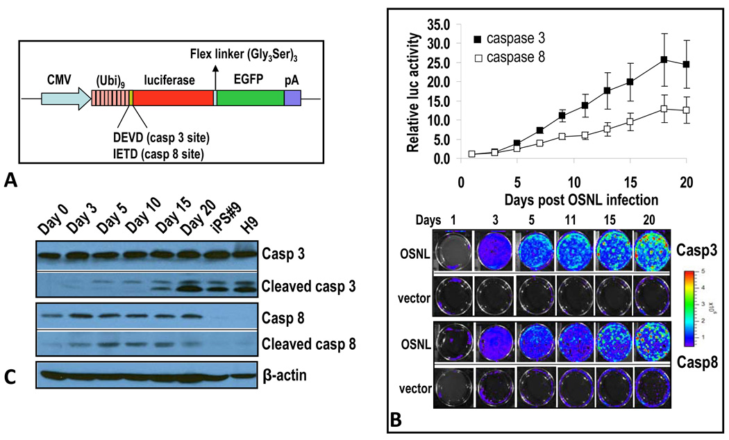
Figure 1. Caspases 3 and 8 are activated in the iPSC induction process.
A) A schematic diagram of caspase reporters based on the proteasome system. Our reporter consists of a fusion reporter domain where a firefly luciferase gene is fused with the enhanced green fluorescence protein (GFP) gene. A polyubiquitin domain is linked to the reporter with caspase 3 (DEVD) and caspase 8 (IETD) cleavages sites. In some cases, the CMV promoter was changed to that of EF1α.
B) Caspase 3 and 8 activation induced by iPSC-inducing transcription factor cocktail. IMR90 cells stably transduced with caspase 3 or caspase 8 reporters were infected with lentiviral vectors encoding OSNL. Reporter activities were then followed through bioluminescence imaging. Top panel, quantitative data showing time-dependent increases in caspase 3 and 8 activities in OSNL-transduced IMR90 cells. Lower panel, representative images of bioluminescence in OSNL-transduced cells. Error bars represent standard error of the mean (SEM, n=3).
C) Western blot analysis of caspase 3 and 8 in IMR90 cells transduced with OSNL. Cells transduced with OSNL were analyzed for total and activated caspase 3 and 8 activities at different times after lentiviral infection. H9 is a commonly used ESC line and iPS#9 is an IMR90-derived iPSC clone. β-actin was used as a protein loading control.
Please also see Figure S1 and Figure S2 for additional related data.

