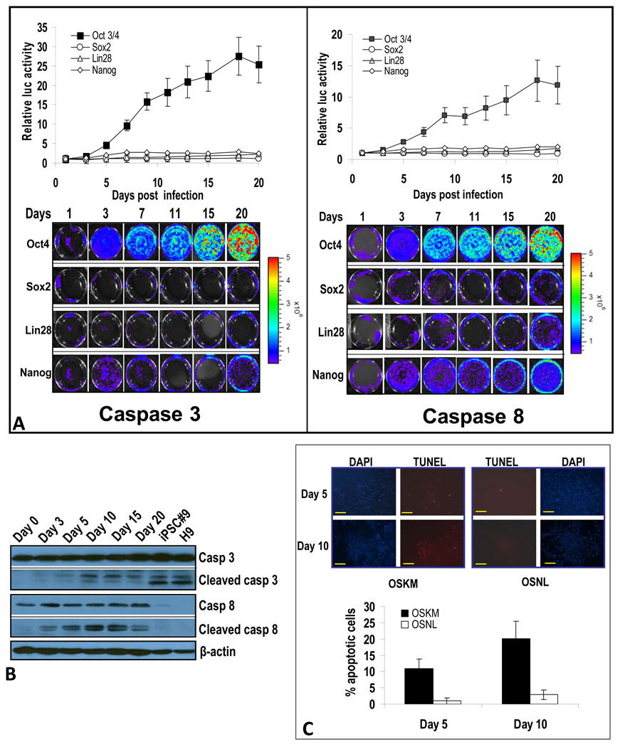
Figure 2. Oct-4 induced activation of caspases 3 and 8 and its effect on fibroblast cell viability.
A) Oct-4 is responsible for activation of caspases 3 and 8 in OSNL-transduced IMR90 cells. Reporter-transduced IMR90 cells were infected with lentiviral vectors encoding individual transcription factors and their reporter activities were followed by bioluminescence imaging. Top panels show the quantitative results of imaging caspases 3 and 8. Lower panels show the bioluminescence imaging of IMR90 cells transduced with caspase 3 and 8 reporters. The error bars represent standard error of the mean (SEM, n=3).
B) Western blot analysis of caspase 3 and 8 activation in Oct-4-transduced IMR90 cells. IMR90 cells infected with Oct-4 encoding lentiviral vectors were examined for caspase 3 and 8 expression and activation at different times after Oct-4 transduction.
C) Cell death in OSNL- and OSKM-transduced cells. IMR90 cells were infected with lentiviral vectors encoding OSNL or OSKM. At five or ten days post transduction, dying cells were quantified through TUNEL staining for DNA fragmentation, which is typical for cells at the end stage of apoptosis. The scale bars represent 200 µm.
Please also see Figure S3 for additional related data.

