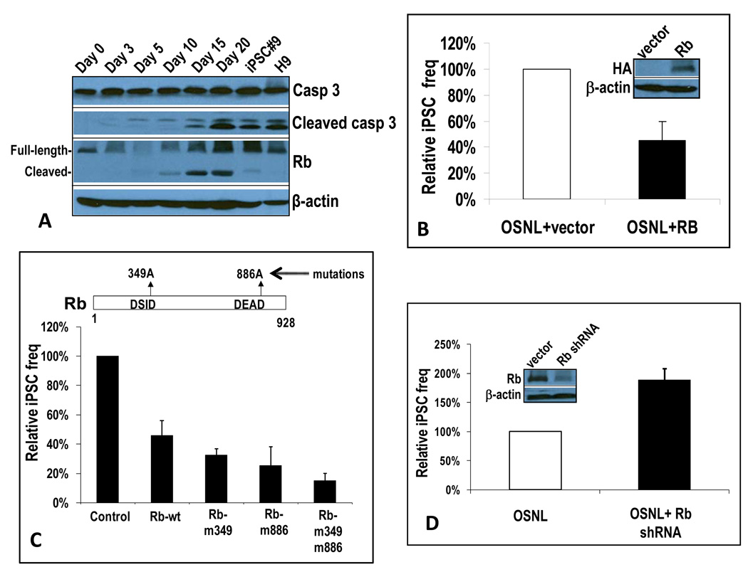
Figure 6. Rb protein as a key downstream target of caspases 3 and 8 in the iPSC induction process.
A) Western blot analysis of Rb expression during the iPSC induction process. IMR90 cells tranaduced with control vector, casp3-DN, or casp8-DN were infected with OSNL or Oct-4. At different time points they were lysed and examined for Rb and other cell cycle regulator/tumor suppressor protein expression through western blot analysis.
B) Rb-mediated suppression of iPSC induction in OSNL-transduced IMR90 cells. A lentiviral vector encoding a full-length Rb gene was co-transduced with OSNL into IMR90 cells. The induction of iPSCs was then examined and quantified. The error bar represents SEM (n=4). Western blots show exogenous Rb expression.
C) The effect of mutations in caspase 3 cleavage sites on the ability of Rb to suppress ONSL induced iPSC formation in IMR90 cells. The error bars represent SEM(n=3).
D) shRNA-mediated knockdown of Rb led to increased iPSC induction in OSNL-transduced IMR90 cells. A lentiviral vector encoding an shRNA minigene targeted to Rb was transduced into IMR90 cells. Cells with stable shRNA expression were then transduced with OSNL and observed for iPSC formation. Relative frequency is shown. The error bar represents SEM (n=4). Western blots show reduced endogenous Rb expression after shRNA transduction .
Please see Figure S6 for additional related data.

